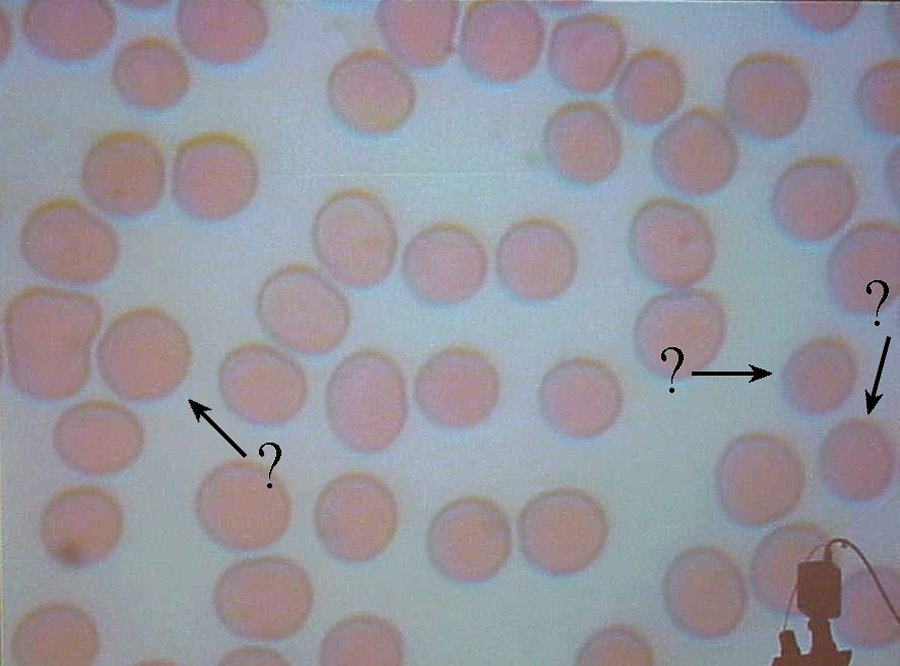
These are amateur photographs made in a general purpose biology lab. They should not be construed as textbook examples.

Discussion: The example slide depicts perhaps one in five or one in six cells is abnormally shaped. My blood is not as severe as that. Still, there are several cells visible which exhibit tendencies in common with Hereditary Spherocytic Hemolytic Anemia. Even given that not all red blood cells are uniformly shaped, the number of cells which appear different in these four images is probably significant enough to suggest the presence of HSHA.
In this image, the two marked cells at the right edge of the frame are substantially smaller and darker than the others which appear on the slide. The marked cell left of center is not darker, but exhibits a very uniform color appearance across its face. Compare this to the darker-and-paler appearing cells below to the right and left, and directly to its right.
 Continue with other thoughts or
return to homepage.