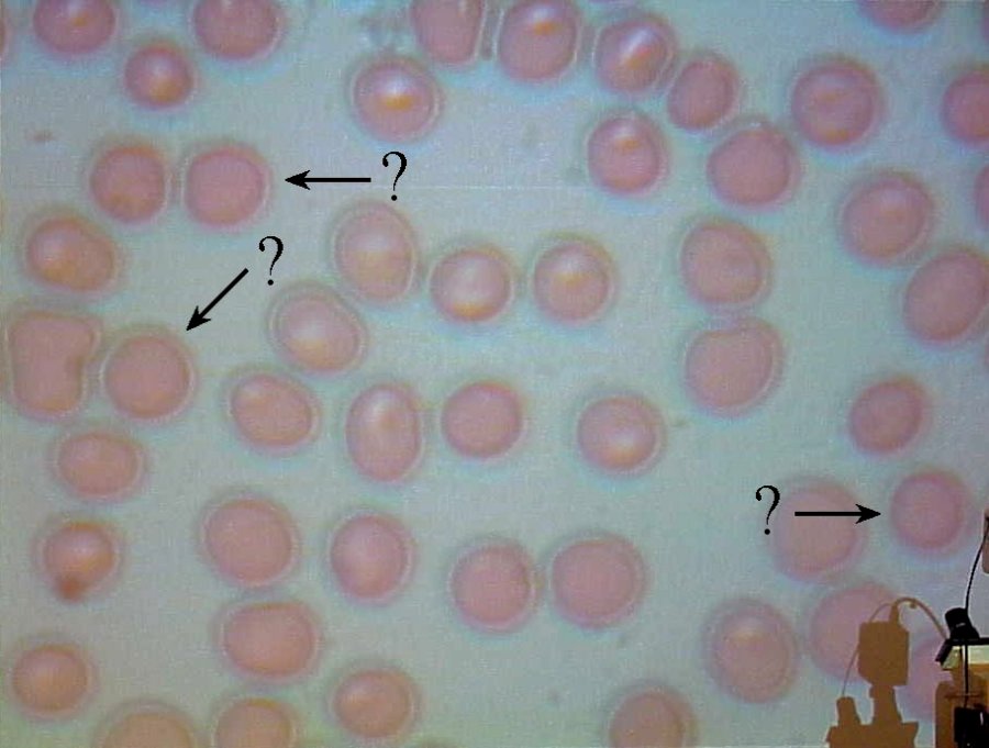
These are amateur photographs made in a general purpose biology lab. They should not be construed as textbook examples.

Discussion: In this image, different levels of light intensity and microscope focal range are used to highlight various aspects of the blood cells' geometries. Notice that the cluster of cells in the center and left of center region have significant highlights in their center regions, showing a good deal of light passage through the cell. In this context, the marked cells in the left third of the image are somewhat darker and noticeably lacking the central region highlights. Also, the marked cell in the lower right corner (near the visible lab equipment) is substantially darker than its neighbors, as well has lacking any center paleness.
 Continue with other thoughts or
return to homepage.